Patholgy Slides : Tumors or Neoplasia
Benign connective tissue tumors
اضغط على الصورة للتكبير -ـ Click on image to enlarge size
Benign tumors of the connective tissue gather all the benign tumors originating from the supportive tissue of various organs and the nonepithelial, extraskeletal structures (exclusive of lymphohematopoietic tissues) : fibrous connective tissue, adipose tissue, skeletal muscle, blood/lymph vessels, and the peripheral nervous system. Benign connective tissue tumors are 100 times more frequent than the malignant ones (sarcomas). Among these, the most frequent are: lipoma, hemangioma, leiomyoma, chondroma and osteoma.
Nomenclature : suffix "oma"+ type of the proliferated tissue.
Nomenclature : suffix "oma"+ type of the proliferated tissue.
Chondroma
Chondroma is a benign cartilaginous tumor, encapsulated, with a lobular growing pattern. Tumor cells (chondrocytes, cartilaginous cells) resemble normal cells and produce the cartilaginous matrix (amorphous, basophilic material). Characteristic are the vascular axes within the tumor, which make the distinction with normal hyaline cartilage. (H&E, ob. x10)
Chondroma. (H&E, ob. x40)
ـــــــــــــــــــــــــــــــــــــــــــــــــــــــــــــــــــــــــــــــــــــــــــــــــــــ
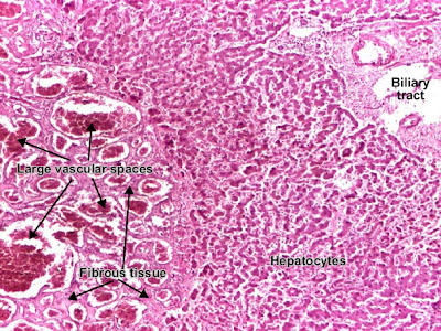
Cavernous hemangioma liver
Cavernous hemangioma is a benign connective tissue tumor resulted from endothelial cells proliferation. It is a non-encapsulated tumor, with an infiltrative, lobular growing. The tumor consists in large (cavernous) spaces, lined by tumor endothelial cells (which appear very similar to normal cells). These interconnected spaces are filled with blood and separated by a fibrous tissue. (H&E, ob. 4)
Cavernous hemangioma (liver). (H&E, ob. X10)
ــــــــــــــــــــــــــــــــــــــــــــــــــــــــــــــــــــــــــــــــــــــــــــــــــــــــــــــ
Leiomyoma (uterus)
Uterine leiomyoma is a benign connective tissue tumor of the smooth muscle cells of the myometrium. Tumor cells resemble normal cells (uniform, elongated, spindle-shaped, with a cigar-shaped nucleus) and form bundles with different directions (whirled). The tumor may present areas of fibrosis, calcification and/or hemorrhage. The tumor is well circumscribed, but not encapsulated. (Hematoxylin-eosin, ob. x10)
ـــــــــــــــــــــــــــــــــــــــــــــــــــــــــــــــــــــــــــــــــــــــــــــــــــــــــــــــــــ

+-+Copy+-+Copy+-+Copy.jpg)


