
Epithelium and Simple Glands
ــــــــــــــــــــــــــــــــــــــــــــــــــــــــــــــــــــــــــ
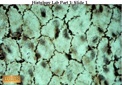
Mesothelium seen as if looking down on a surface view to see "pavement" effect of the lining cells. Silver stains the intercellular cement dark between adjacent cells. Notice how corrugated the cell membranes are. Mesothelium = the simple squamous epithelium lining body cavities and mesenteries.
ـــــــــــــــــــــــــــــــــــــــــــــــــــــــــــــــــــــــــــــــــــــــــــــــــــــــــــــــــــــــــــــــــــــــــــــــــــــــــــــــــــ
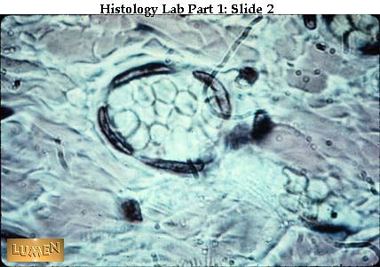
High power view of endothelial cells lining a small blood vessel cut in cross-section. (You see just the nuclei - the cytoplasm between them is extremely flat.) Endothelium = the simple squamous epithelium lining blood vessels.
ـــــــــــــــــــــــــــــــــــــــــــــــــــــــــــــــــــــــــــــــــــــــــــــــــــــــــــــــــــــــــــــــــــــــــــــــــــــــــــــــــ
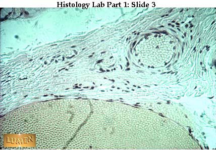
Low power view of larger vessels, showing endothelial nuclei lining the lumen. The yellowish cells filling each vessel's lumen are blood cells.
ـــــــــــــــــــــــــــــــــــــــــــــــــــــــــــــــــــــــــــــــــــــــــــــــــــــــــــــــــــــــــــــــــــــــــــــــــــــــــــــــ
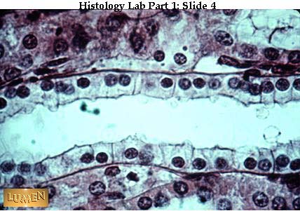
Simple cuboidal epithelium lining a tubule (longitudinal cut). Some of the cell boundaries between "blocks" or "cubes" here are quite distinct.
ــــــــــــــــــــــــــــــــــــــــــــــــــــــــــــــــــــــــــــــــــــــــــــــــــــــــــــــــــــــــــــــــــــــــــــــــــــــــــــ
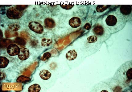
Simple cuboidal epithelium in Mallory stain (longitudinal cut). Note the dark chromatin clumps in the nuclei. Underneath the epithelium lies a small blood vessel filled with orange-colored blood cells.
ــــــــــــــــــــــــــــــــــــــــــــــــــــــــــــــــــــــــــــــــــــــــــــــــــــــــــــــــــــــــــــــــــــــــــــــــــــــــــــــــــ
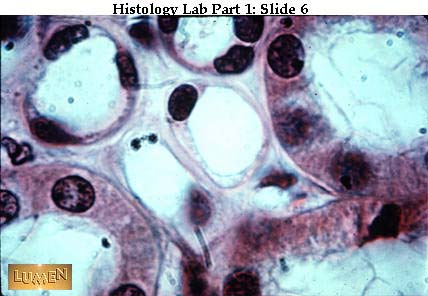
Cross-section of tubules. The smaller ones clustered in the center and upper left are lined by simple squamous epithelium. The larger pink tubules have simple cuboidal epithelium.
ـــــــــــــــــــــــــــــــــــــــــــــــــــــــــــــــــــــــــــــــــــــــــــــــــــــــــــــــــــــــــــــــــــــــــــــــــــــــــــــــــــــــ

A tubule stained to show the pink basement membrane underlying the base of the simple cuboidal epithelium. Stained with periodic acid Schiff reagent (PAS), which stains mucopolysaccharides.
ـــــــــــــــــــــــــــــــــــــــــــــــــــــــــــــــــــــــــــــــــــــــــــــــــــــــــــــــــــــــــــــــــــــــــــــــــــــــــــــــــــ
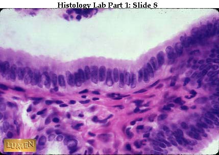
Simple columnar epithelium with very regular line-up of nuclei.
ـــــــــــــــــــــــــــــــــــــــــــــــــــــــــــــــــــــــــــــــــــــــــــــــــــــــــــــــــــــــــــــــــــــــــــــــــــــــــــــــــــــــــــــــ
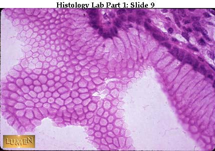
Simple columnar cells cut tangentially to show how they form a very regular "pavement" when viewed from the surface. The cells are like tall blocks arranged very closely to each other with a small amount of tissue fluid in between.
ــــــــــــــــــــــــــــــــــــــــــــــــــــــــــــــــــــــــــــــــــــــــــــــــــــــــــــــــــــــــــــــــــــــــــــــــــــــــــــ
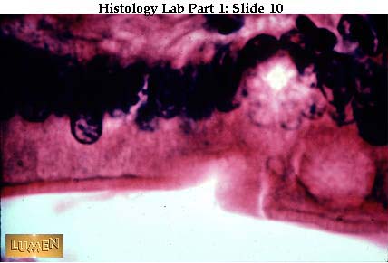
Detail of simple columnar epithelium with striated border (microvilli). Notice that the border is quite thin and the striations close together, looking like very regular, closely set brush bristles.
ــــــــــــــــــــــــــــــــــــــــــــــــــــــــــــــــــــــــــــــــــــــــــــــــــــــــــــــــــــــــــــــــــــــــــــــــــــــــــــــــــــــــــ
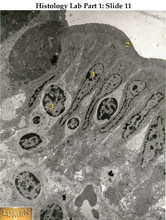
EM of cells with striated border. Notice the evenness and regularity of the microvilli. This is an adaptation of the cell surface for absorption. Notice also the corrugation of the cell boundaries as they fit next to each other. 1= nucleus; 2=brush border (microvilli); 3=lymphocyte.
ــــــــــــــــــــــــــــــــــــــــــــــــــــــــــــــــــــــــــــــــــــــــــــــــــــــــــــــــــــــــــــــــــــــــــــــــــــــــــــــــ
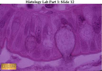
Detail of simple columnar epithelium with a goblet cell secreting mucus. The thin, clearly defined band along the top epithelial surface is the striated border, though the individual striations (or microvilli) are not visible at this magnification. The lower edge of the striated border is the location of the terminal web; the dots along the line of the web, seen in between the individual epithelial cells, are the so-called terminal bars, which are found in EM to consist of various cell junctions.
ـــــــــــــــــــــــــــــــــــــــــــــــــــــــــــــــــــــــــــــــــــــــــــــــــــــــــــــــــــــــــــــــــــــــــــــــــــــــــــــــــــ
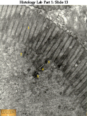
EM of apical (top) surface of two epithelial cells whose cell membranes lie next to each other. The microvilli (1) of the striated border are very straight and regimented in appearance. Microfilaments within them can be seen extending down into the terminal web (2), which is an aggregate of fine filaments lying in the cell cytoplasm. Several junctional complexes are seen including tight junction (zonula occludens =3); intermediate junction (zonula adherens =4); and desmosome (macula adherens =5).
ــــــــــــــــــــــــــــــــــــــــــــــــــــــــــــــــــــــــــــــــــــــــــــــــــــــــــــــــــــــــــــــــــــــــــــــــــــــــــــــــــــــــ
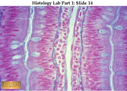
Four rows of simple columnar epithelium facing each other in pairs (left and right) across a narrow lumen or channel that lies in the middle of each pair. (This is a Mallory Trichrome stain.) The goblet cells are filled with blue mucoid secretion which is being poured into the narrow lumens. Notice that in all four rows of epithelium there is a narrow band of striated border next to the lumen; the dark purple line at the base of the border is the terminal web. Look at the right hand rows of epithelial cells and notice the dark dots all along the terminal web lines; these dots represent the junctional complexes between cells. The central cavity in the picture is a blood vessel with endothelium, surrounded by a very cellular connective tissue. Separating this connective tissue from the epithelium is a thin blue layer of connective tissue fibers.
ــــــــــــــــــــــــــــــــــــــــــــــــــــــــــــــــــــــــــــــــــــــــــــــــــــــــــــــــــــــــــــــــــــــــــــــــــــــــــــــــــــــــــــ
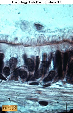
Pseudostratified ciliated columnar epithelium from the trachea. Nuclei are at different levels. All cells touch the basement membrane, but only the taller cells reach the lumen. The cilia are longer and less regular than the microvilli of a striated border.
ـــــــــــــــــــــــــــــــــــــــــــــــــــــــــــــــــــــــــــــــــــــــــــــــــــــــــــــــــــــــــــــــــــــــــــــــــــــــــــــــــــ
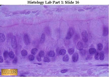
Pseudostratified ciliated columnar epithelium with pale goblet cells. The different levels of nuclei are clearer here. Again, notice the wavy-looking cilia.
ـــــــــــــــــــــــــــــــــــــــــــــــــــــــــــــــــــــــــــــــــــــــــــــــــــــــــــــــــــــــــــــــــــــــــــــــــــــــــــــــــــــــــــ
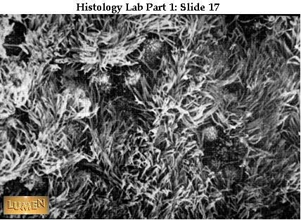
Surface view of cilia with scanning EM scope. Notice how "ragged" the surface seems -- cilia were caught as they moved.
ـــــــــــــــــــــــــــــــــــــــــــــــــــــــــــــــــــــــــــــــــــــــــــــــــــــــــــــــــــــــــــــــــــــــــــــــــــــــــــــــــــــ
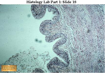
Transitional epithelium of the urinary bladder, low power view. It is a stratified epithelium with several layers of cells.
ــــــــــــــــــــــــــــــــــــــــــــــــــــــــــــــــــــــــــــــــــــــــــــــــــــــــــــــــــــــــــــــــــــــــــــــــــــــــــــــــــــــــــــــــ
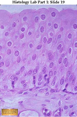
Transitional epithelium, high power. Notice many layers of cells -- and the typically puffy surface cells. The bladder is contracted so the epithelium is thick. If the bladder were stretched, the epithelium would be thinner.
ــــــــــــــــــــــــــــــــــــــــــــــــــــــــــــــــــــــــــــــــــــــــــــــــــــــــــــــــــــــــــــــــــــــــــــــــــــــــــــــــــــ
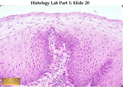
Stratified squamous non-cornified epithelium -- medium power. This is from the esophagus, so the surface is moist and living. Surface cells are squamous and still nucleated. Basal layer is very distinct; compare this with the less distinct basal layer of the preceding slide of transitional epithelium.
ــــــــــــــــــــــــــــــــــــــــــــــــــــــــــــــــــــــــــــــــــــــــــــــــــــــــــــــــــــــــــــــــــــــــــــــــــــــــــــــــــــــــــــــ
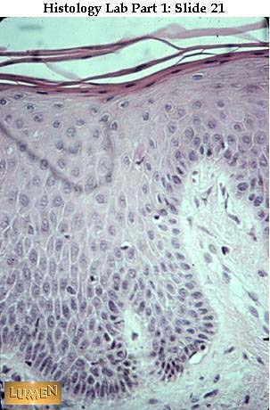
Stratified squamous epithelium with beginning surface cornification. This section is from thin skin, which has a dry surface covered with dead cells. Notice how flat the surface cells are and how dark and pyknotic their nuclei have become. Again, notice the distinct row of basal cells.
ـــــــــــــــــــــــــــــــــــــــــــــــــــــــــــــــــــــــــــــــــــــــــــــــــــــــــــــــــــــــــــــــــــــــــــــــــــــــــــــــــــــــــ
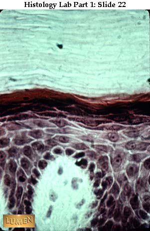
Thickly cornified stratified squamous epithelium. The cells in the bright red layer and in the pale layers above it are completely flattened and dead, and have lost their nuclei.
ـــــــــــــــــــــــــــــــــــــــــــــــــــــــــــــــــــــــــــــــــــــــــــــــــــــــــــــــــــــــــــــــــــــــــــــــــــــــــــــــــ
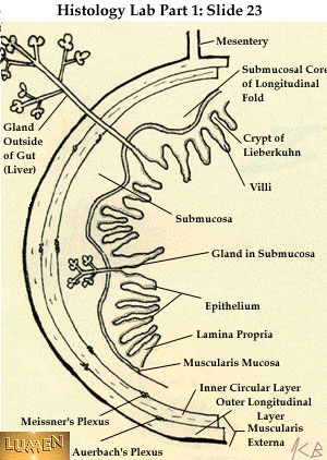
Diagram of GI wall to show various kinds of glands -- some within the wall and some without (like the liver). These glands have ducts that empty into the lumen of the gut. In all cases, the epithelium lining the ducts and glands is continuous with the epithelium lining the lumen (cavity) of the gut. (Note: the test-tube-like glands, labeled "crypts of Lieberkuhn" here, are the same kind as the intestinal glands you saw under the microscope in the appendix in lab.)
ــــــــــــــــــــــــــــــــــــــــــــــــــــــــــــــــــــــــــــــــــــــــــــــــــــــــــــــــــــــــــــــــــــــــــــــــــــــــــــــــــــــــــــ
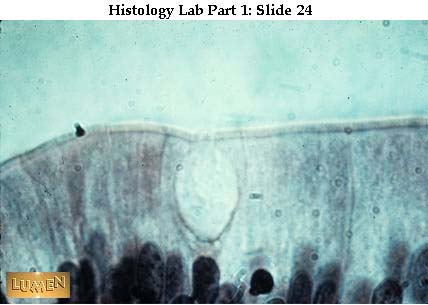
Unicellular gland - a goblet cell mucus-secreting. (H & E stain.)
ــــــــــــــــــــــــــــــــــــــــــــــــــــــــــــــــــــــــــــــــــــــــــــــــــــــــــــــــــــــــــــــــــــــــــــــــــــــــــــــــــــــــ

Goblet cells (blue) scattered throughout simple columnar epithelial lining (special quad stain).
ــــــــــــــــــــــــــــــــــــــــــــــــــــــــــــــــــــــــــــــــــــــــــــــــــــــــــــــــــــــــــــــــــــــــــــــــــــــــــــــــــــــــــــــــــ

Simple tubular glands of gut wall seen in low power. These glands are lined with epithelium throughout their whole extent.
ـــــــــــــــــــــــــــــــــــــــــــــــــــــــــــــــــــــــــــــــــــــــــــــــــــــــــــــــــــــــــــــــــــــــــــــــــــــــــــــــــــــــــ
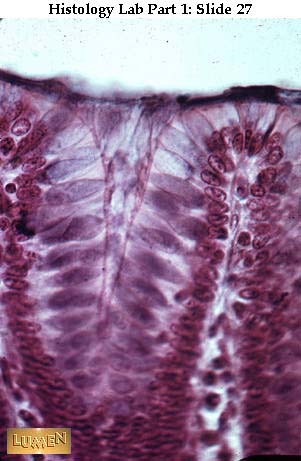
Detail of such a gland. Goblet cells are purple here. -(H & E)
ـــــــــــــــــــــــــــــــــــــــــــــــــــــــــــــــــــــــــــــــــــــــــــــــــــــــــــــــــــــــــــــــــــــــــــــــــــــــــــــــــــــــــــــــــــ
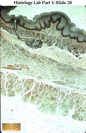
Low power of wall of esophagus showing duct at right, leading down to simple tubulo-alveolar gland with coiled secretory portions.
ــــــــــــــــــــــــــــــــــــــــــــــــــــــــــــــــــــــــــــــــــــــــــــــــــــــــــــــــــــــــــــــــــــــــــــــــــــــــــ
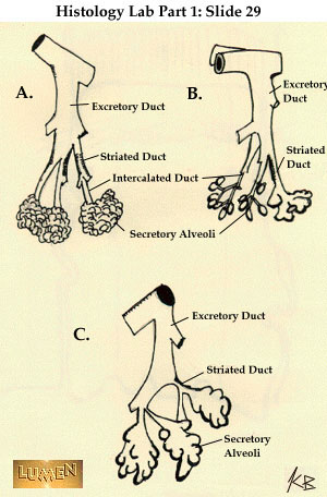
Drawings of compound tubulo-alveolar glands -- showing the branching of their duct system -- and a few secretory end-pieces (alveoli). Ducts and alveoli are lined with epithelium.
ــــــــــــــــــــــــــــــــــــــــــــــــــــــــــــــــــــــــــــــــــــــــــــــــــــــــــــــــــــــــــــــــــــــــــــــــــــــــــــــــــــــــــــــــ
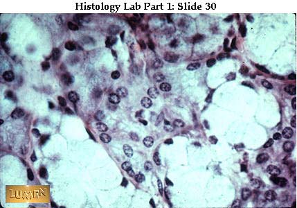
High power of typical mucous (pale) and serous (darker pink) secretory cells. Notice that the nuclei of mucous cells are dark and flattened at the base of the cells, while the nuclei of serous cells are round and more centrally located at their cells. Mucous secretion is relatively thick and viscous; serous secretion is watery.
ــــــــــــــــــــــــــــــــــــــــــــــــــــــــــــــــــــــــــــــــــــــــــــــــــــــــــــــــــــــــــــــــــــــــــــــــــــــــــــــــــــــــــــــــــــــــــــــــــــــ
