
+-+Copy+-+Copy+-+Copy.jpg)
+-+Copy+-+Copy+-+Copy.jpg)
Patholgy Slides : Tumors or Neoplasia
Benign epithelial tumors
اضغط على الصورة للتكبير - Click on image to enlarge size
Squamous cell papilloma (skin)

Squamous cell papilloma (verruca vulgaris) is a benign epithelial tumor. Tumor cells proliferate and produce finger-like or warty projections; secondary, the subjacent vessels and connective tissue (fibrovascular core) grows to sustain and feed the tumor. The tumor cells resemble normal squamous cells, but there is an increase of the layers number: acanthosis, hypergranulosis and hyperkeratosis. The basement membrane is intact. (H&E, ob. X10)
ــــــــــــــــــــــــــــــــــــــــــــــــــــــــــــــــــــــــــ
 Adenomatous tubulo-villous polyp, pedunculated (colon).
The adenomatous proliferation is characterized by different degrees of cell dysplasia (cellular and architectural atypia) : loss of normal differentiation of epithelium, irregular cells with hyperchromatic nuclei, (pseudo)stratified nuclei, nucleolus, decreased mucosecretion and mitosis. The architecture may be tubular, villous or tubulo-villous. Basement membrane and muscularis mucosae are intact. (HE, ob. x4)
Detail of an adenomatous tubulo-villous polyp (colon)
 Compared to normal colonic epithelium (lower left corner of the picture) the adenomatous proliferation is characterized by different degrees of cell dysplasia and tubular, villous or tubulo-villous architecture. In tubular component tumor cells proliferate generating tubular shapes, more or less irregular or branched. The villous pattern presents villi - finger-like projections lined by dysplastic epithelium and with thin fibro-vascular core. Passage from tumoral epithelium to normal mucosa is abrupt. (HE, ob. x10)
ـــــــــــــــــــــــــــــــــــــــــــــــــــــــــــــــــــــــــــ
In hollow organs (e.g. digestive tract) the adenoma grows upwards into the lumen - adenomatous polyp or polypoid adenoma. Depending on the type of the insertion base, adenoma may be pedunculated (lobular head with a long, slender stalk, covered by normal mucosa) or sessile (broad base - see photo). If not elevated above the surface of the mucosa, the adenoma is called flat adenoma
 Adenomatous villous polyp, sessile (colon). The adenomatous proliferation is characterized by different degrees of cell dysplasia (cellular and architectural atypia): loss of normal differentiation of epithelium, irregular cells with hyperchromatic nuclei, (pseudo)stratified nuclei, nucleolus, decreased mucosecretion and atypical mitosis. The architecture may be tubular, villous (photo) or tubulo-villous. Basement membrane and muscularis mucosae are intact. (HE, ob. x4)
ــــــــــــــــــــــــــــــــــــــــــــــــــــــــــــــــــــــــــ
Fibroadenoma of the breast

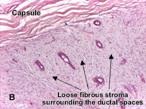 Fibroadenoma of the breast is a benign tumor composed of two elements : epithelium and stroma. It is nodular and encapsulated, included in breast. The epithelial proliferation appears in a single terminal ductal unit and describes duct-like spaces surrounded by a fibroblastic stroma. Depending on the proportion and the relationship between these two components, there are two main histological features : intracanalicular and pericanalicular. Often, both types are found in the same tumor. Intracanalicular fibroadenoma (photo A) : stromal proliferation predominates and compresses the ducts, which are irregular, reduced to slits. Pericanalicular fibroadenoma (photo B) : fibrous stroma proliferates around the ductal spaces, so that they remain round or oval, on cross section. The basement membrane is intact. (H&E, ob. x10)
ـــــــــــــــــــــــــــــــــــــــــــــــــــــــــــــــــــــــــــــــــــــــــــ
ــــــــــــــــــــــــــــــــــــــــــــــــــــــــــــــــــــــــــ
Adenomatous tubulo-villous polyp, pedunculated (colon)
Adenoma is a benign epithelial tumor arising in epithelium of mucosa (stomach, small intestine and bowel), glands (endocrine and exocrine) and ducts. In hollow organs (e.g. digestive tract) the adenoma grows upwards into the lumen - adenomatous polyp or polypoid adenoma. Depending on the type of the insertion base, adenoma may be pedunculated (lobular head with a long, slender stalk, covered by normal mucosa - see photo) or sessile (broad base). If not elevated above the surface of the mucosa, the adenoma is called flat adenoma
The adenomatous proliferation is characterized by different degrees of cell dysplasia (cellular and architectural atypia) : loss of normal differentiation of epithelium, irregular cells with hyperchromatic nuclei, (pseudo)stratified nuclei, nucleolus, decreased mucosecretion and mitosis. The architecture may be tubular, villous or tubulo-villous. Basement membrane and muscularis mucosae are intact. (HE, ob. x4)
Detail of an adenomatous tubulo-villous polyp (colon)

ـــــــــــــــــــــــــــــــــــــــــــــــــــــــــــــــــــــــــــ
Adenomatous villous polyp, sessile (colon)
Adenoma is a benign epithelial tumor arising in epithelium of mucosa (stomach, small intestine and bowel), glands (endocrine and exocrine) and ducts.In hollow organs (e.g. digestive tract) the adenoma grows upwards into the lumen - adenomatous polyp or polypoid adenoma. Depending on the type of the insertion base, adenoma may be pedunculated (lobular head with a long, slender stalk, covered by normal mucosa) or sessile (broad base - see photo). If not elevated above the surface of the mucosa, the adenoma is called flat adenoma

ــــــــــــــــــــــــــــــــــــــــــــــــــــــــــــــــــــــــــ
Fibroadenoma of the breast


ـــــــــــــــــــــــــــــــــــــــــــــــــــــــــــــــــــــــــــــــــــــــــــ